Lungs And Diseases
Understanding Lungs and Their Diseases: An Educational Overview
Welcome to this session presented by Saffron Educational and Medical Foundation. In today’s session, we focus on understanding the human lungs—their structure, function, common diseases, causes, and natural treatment approaches including naturopathy, diet, diagnostic methods, and yoga therapy.

Introduction to the Lungs
The human body contains two lungs—right and left—located in the chest cavity. Together, these two organs occupy a large portion of the intrathoracic (chest) space. Their primary function is gas exchange: supplying oxygen to the bloodstream while removing carbon dioxide.
Basic Anatomy and Function
- Right and Left Lungs: Positioned in the thoracic cavity.
- Pleural Cavity: Each lung is encased in a membrane called the pleura, which creates a protective cavity.
- Main Bronchus and Trachea: The trachea (windpipe) divides into the main bronchi, connecting both lungs to the upper respiratory system.
- Pulmonary Arteries and Heart: The lungs work closely with the heart via pulmonary arteries, ensuring blood is oxygenated.
The main purpose of this structure is to facilitate respiration, making oxygen available to body tissues and removing waste gases.
Components of the Respiratory System

The human respiratory system is made up of several key parts:
- Nasal Cavity and Nose: The entry point for air.
- Pharynx and Larynx: Passageways that guide air towards the lungs.
- Trachea: The windpipe that branches into the bronchi.
- Bronchi and Bronchioles: Airways that lead air directly into the lungs.
- Lungs: The central organs for gas exchange.
- Alveoli: Small air sacs where actual gas exchange occurs.
This sequence ensures air passes through a clean, moistened, and temperature-regulated pathway before reaching the lungs.
Lung Functions Beyond Breathing
Though primarily responsible for breathing, the lungs also:
- Help regulate the body’s pH balance.
- Filter out small blood clots.
- Provide a defense against inhaled pathogens and particles.
Common Lung Diseases
Lung diseases can result from infections, environmental factors, lifestyle choices, or genetic predisposition. Here are key examples:
- Asthma: Chronic inflammation of airways causing wheezing and shortness of breath.
- Chronic Obstructive Pulmonary Disease (COPD): Includes emphysema and chronic bronchitis.
- Pneumonia: Infection causing inflammation in the air sacs.
- Tuberculosis (TB): A serious infectious disease affecting the lungs.
- Lung Cancer: Malignant tumors in the lung tissues.
- Pulmonary Fibrosis: Scarring of lung tissue leading to breathing difficulties.
Causes of Lung Diseases
- Environmental Factors: Pollution, allergens, occupational hazards.
- Lifestyle Choices: Smoking, poor diet, lack of exercise.
- Infections: Bacterial, viral, or fungal infections.
- Genetic Predisposition: Family history of lung diseases.
Naturopathic and Food-Based Treatments
Natural approaches emphasize prevention, holistic treatment, and support through lifestyle changes:
- Diet:
- Include antioxidant-rich foods like berries, green leafy vegetables, and nuts.
- Stay hydrated to support mucous membrane function.
- Reduce inflammatory foods such as processed sugars and trans fats.
- Naturopathy Therapies:
- Steam Inhalation: Helps open airways and clear congestion.
- Hydrotherapy: Alternate hot and cold water therapy may support lung function.
- Herbal Treatments: Using natural herbs such as licorice root, ginger, and turmeric.
- Yoga Therapy:
Certain yoga postures and breathing techniques (Pranayama) improve lung capacity and oxygen intake:- Anulom Vilom (Alternate Nostril Breathing)
- Kapalbhati (Skull Shining Breath)
- Bhastrika (Bellows Breath)
Diagnostic Methods for Lung Diseases
- Chest X-rays and CT Scans: Visual imaging of lung structures.
- Pulmonary Function Tests (PFTs): Measure lung capacity and airflow.
- Sputum Tests: Identify infections or abnormal cells.
- Oxygen Saturation Monitoring: Measure oxygen levels in the blood.
Practical Lifestyle Recommendations
- Avoid smoking or exposure to secondhand smoke.
- Exercise regularly with focus on cardiovascular fitness.
- Maintain proper posture to support lung expansion.
- Practice daily breathing exercises.
- Ensure adequate rest and hydration.
Detailed Anatomy of the Lungs: Lobes, Fissures, and Functional Zones

The Hilum: Gateway of the Lungs
At the medial side of each lung, there is a specialized region called the hilum. The hilum serves as the point through which major structures enter and exit the lungs, including:
- Pulmonary arteries: Carry deoxygenated blood from the heart to the lungs.
- Pulmonary veins: Carry oxygenated blood back to the heart.
- Bronchi: Main airways into the lungs.
- Lymphatic vessels and nerves: Important for immune defense and nervous system control.
The hilum is essential for maintaining both respiratory and circulatory functions, acting as a critical hub for all vascular and nervous connections.
Lobes of the Lungs
Humans have two lungs, but they are not symmetrical in size or structure due to the positioning of the heart.
- Right Lung:
- Three Lobes: Superior, Middle, and Inferior Lobes.
- Larger in Size: Slightly larger than the left lung because the heart tilts towards the left.
- Two Fissures:
- Oblique Fissure: Separates the middle lobe from the inferior lobe.
- Horizontal Fissure: Separates the superior lobe from the middle lobe.
- Left Lung:
- Two Lobes: Superior and Inferior Lobes.
- Smaller in Size: Due to the heart’s leftward tilt creating a visible notch known as the cardiac notch.
- One Fissure:
- Oblique Fissure: Divides the two lobes.
Why the Size Difference?
The left lung’s smaller size is an evolutionary adaptation to accommodate the heart’s positioning within the chest cavity. This anatomical feature is called the cardiac notch and is clearly visible in anatomical diagrams.
Functional Zones Within the Lungs
- Apex of the Lung:
The uppermost portion of each lung, located near the clavicle (collarbone). - Base of the Lung:
The bottom part of the lung that rests on the diaphragm. - Costal Surface:
The side of the lungs adjacent to the ribs. - Diaphragmatic Surface:
The surface of the lungs in contact with the diaphragm, playing a vital role in breathing movements.
The Trachea: Structure, Function, and Its Role in Respiratory Health
Basic Anatomy and Positioning
The trachea, commonly referred to as the windpipe, is a vital structure in the respiratory system. It is positioned:
- Below the larynx (voice box)
- In between the two lungs
- In front of the esophagus (food pipe)
The trachea serves as the primary airway, conducting air from the nasal cavity down towards the bronchi and lungs.
Structural Characteristics of the Trachea

- Lined with Mucous Membrane:
- The trachea is internally lined with a specialized mucous membrane.
- This lining produces mucus, which plays a crucial role in trapping dust, microbes, and other foreign particles that enter the respiratory system.
- Supported by Cartilaginous Rings:
- The trachea is structured with C-shaped cartilage rings.
- These rings provide:
- Rigidity and structure: Preventing the windpipe from collapsing.
- Flexibility: Allowing slight movement and flexibility during breathing and swallowing.
- The open part of the “C” faces the esophagus, allowing it to expand when swallowing food.
- Relationship with the Esophagus:
- The esophagus lies directly behind the trachea.
- The C-shaped cartilage design accommodates the esophagus, ensuring that swallowing does not obstruct the airway.
Function of the Tracheal Mucus
The mucous membrane lining the trachea plays a vital protective role:
- Traps Foreign Particles: Dust, allergens, pathogens, and other foreign substances are trapped by the mucus.
- Cilia Movement: Tiny hair-like structures called cilia line the trachea and move the mucus upwards towards the throat.
- Removal of Contaminants:
- The mucus, along with the trapped particles, is either:
- Swallowed and neutralized in the stomach.
- Expelled through coughing or sneezing.
- The mucus, along with the trapped particles, is either:
This mechanism is known as the mucociliary escalator, and it is crucial for maintaining lung cleanliness and preventing infections.
Pulmonary Segments, Bronchi, and Bronchioles: An Essential Overview of Lung Anatomy
Pulmonary Segments: The Building Blocks Within Lung Lobes
The lungs are divided into lobes — three on the right and two on the left — but the division doesn’t end there. Each lobe is further subdivided into smaller functional units known as pulmonary segments.
- Right Lung: Typically has 10 pulmonary segments.
- Left Lung: Can have 8 to 10 pulmonary segments depending on anatomical classification.
Why Pulmonary Segments Matter:
- Each segment functions independently.
- Segments have their own bronchus, artery, and vein.
- Clinically, knowing these segments is vital for procedures like surgery, bronchoscopy, or segmental resections.
The Bronchi: The Primary Airways
Once air passes through the trachea, it enters the bronchi.
Structure and Division:
- The trachea splits into two main branches at its lower end:
- Right main bronchus (wider and shorter).
- Left main bronchus (narrower and longer).
Inverted Y-Shaped Division:
- This splitting forms an inverted Y shape.
- The bronchi are surrounded by irregular cartilage plates instead of complete rings, providing flexibility while maintaining airway patency.
Further Division:
The bronchi branch progressively:
- Primary Bronchi → Main branches from the trachea.
- Secondary Bronchi → Supply each lobe of the lung.
- Tertiary Bronchi (Segmental Bronchi) → Supply each pulmonary segment.
- Bronchioles → Smaller airways branching from the bronchi.
Bronchioles: Conducting Airways Without Cartilage
Key Characteristics:
- Bronchioles lack cartilage.
- Unlike bronchi, bronchioles are not supported by cartilage plates or rings.
- Lined with simple columnar or cuboidal epithelium.
- This type of lining helps with the smooth passage of air while offering some filtration.
- Surrounded by smooth muscle fibers.
- Bronchioles can constrict or dilate, regulating airflow resistance and volume.
Functional Importance:
- Bronchioles are considered conducting airways.
- They deliver air to alveoli — the tiny air sacs where gas exchange occurs.
- The absence of cartilage makes bronchioles more flexible but also more vulnerable to collapse, especially in conditions like asthma or bronchitis.
Alveoli: The Functional Units of Gas Exchange in the Lungs

What Are Alveoli?
At the end of the bronchial tree, bronchioles terminate in tiny, spongy sacs called alveoli.
- Singular form: Alveolus
- Alveoli represent the primary site of gas exchange in the lungs.
Structure and Function:
- Air-filled sacs surrounded by a dense network of capillaries (tiny blood vessels).
- Each lung contains approximately 300 million alveoli, increasing the surface area for gas exchange.
- The walls of alveoli are extremely thin and permeable, allowing oxygen and carbon dioxide to pass through easily.
The Gas Exchange Process:
- Inhaled air reaches the alveoli.
- Oxygen passes through alveolar walls into the capillary blood.
- Carbon dioxide moves from the blood into the alveoli to be exhaled.
- The oxygen-rich blood circulates through the body, supplying tissues and organs.
This is how the lungs oxygenate the blood and remove carbon dioxide waste efficiently.
The Diaphragm: The Muscle That Powers Breathing
Location and Structure:
- The diaphragm is a strong, dome-shaped sheet of muscle located at the base of the lungs.
- It forms a partition between:
- The thoracic cavity (chest) — where the lungs and heart are located.
- The abdominal cavity — housing digestive organs.
How the Diaphragm Works:
- During inhalation:
- The diaphragm contracts and flattens, creating more space in the chest cavity.
- This reduces pressure inside the lungs, allowing air to flow in.
- During exhalation:
- The diaphragm relaxes and returns to its dome shape.
- This increases pressure, helping push air out of the lungs.
Why It Matters:
- The diaphragm is crucial for breathing and maintaining the rhythmic airflow required for life.
- It’s an involuntary muscle — controlled automatically by the respiratory center in the brain, although you can consciously control it (like holding your breath).
The Role of the Diaphragm in Breathing
How the Diaphragm Supports Lung Function
- Contraction and Relaxation:
- When the diaphragm contracts, it moves downward, expanding the chest cavity and allowing the lungs to fill with air (inhalation).
- When the diaphragm relaxes, it moves upward, reducing the chest cavity’s volume and helping push air out (exhalation).
- Feel the Movement:
- By placing your hand on your chest or abdomen, you can physically feel the expansion and relaxation cycle as you breathe in and out.
- This is one complete respiratory cycle.
Gross Anatomy of the Lungs
Basic Characteristics:
- Texture: Spongy
- Color: Pinkish-grey in healthy lungs
- Shape: Cone-shaped, each with an apex (top) and base (bottom)
Surfaces of the Lungs:
- Costal Surface:
- Faces the ribs (costal = ribs)
- Joins with the mediastinal surface anteriorly
- Mediastinal Surface:
- Faces the mediastinum
- Where important structures like the heart, pericardium, blood vessels, and airways connect to the lungs
- Diaphragmatic Surface:
- Concave
- Rests on the diaphragm
Borders of the Lungs:
- Anterior Border
- Posterior Border
- Inferior Border
Special Anatomical Features

Cardiac Notch
- Location: Present only in the left lung
- Purpose:
- Accommodates the heart’s left ventricle
- Creates a distinct indentation along the anterior border
Lingula
- Location: Also part of the left lung
- Function:
- A small projection below the cardiac notch
- Balances lung volume since the right lung is larger
Dome of the Diaphragm
- Right Dome: Higher due to the liver underneath.
- Left Dome: Slightly lower.
Detailed Anatomy of the Lungs: Borders, Lobes, and Segments
Lung Borders and Landmarks:
- Inferior Border
- Thin and separates the lung base from the costal surface.
- Important for identifying lung positioning in medical imaging.
- Posterior Border
- Thicker than the inferior border.
- Extends from cervical vertebra C7 down to thoracic vertebra T10.
- This region is palpable, especially at C7 (noted as a prominent vertebral bump on the back of the neck).
Bronchial Tree Hierarchy:

- Primary Bronchi
- First branch from the trachea.
- Secondary (Lobar) Bronchi
- Branches from the primary bronchi to each lobe.
- Tertiary (Segmental) Bronchi
- Branches from the secondary bronchi.
- Supply specific bronchopulmonary segments.
- Right lung: 10 segments.
- Left lung: 8–10 segments.
Mnemonic for Right Lung Segments:
In anatomy teaching, mnemonics are often used. The transcript mentioned a mnemonic but didn’t spell it out. A commonly taught mnemonic for the right lung segments is:
- Apical
- Posterior
- Anterior
- Lateral
- Medial
- Superior
- Medial Basal
- Anterior Basal
- Lateral Basal
- Posterior Basal
Right Lung Segment Summary:
- Upper Lobe Segments (3):
- Apical (top)
- Posterior (back)
- Anterior (front)
- Middle Lobe Segments (2):
- Lateral segment
- Medial segment (closer to the heart)
- Lower Lobe Segments (5):
- Superior segment
- Medial basal segment
- Lateral basal segment
- Anterior basal segment
- Posterior basal segment
- Mnemonic Suggestion (Right Lung Segments):
- “APe’s ALPS MLaP”
- Apical, Posterior, Anterior
- Lateral, Medial
- Superior, Medial Basal, Lateral Basal, Anterior Basal, Posterior Basal
- “APe’s ALPS MLaP”
Left Lung Segment Summary:

- 8–10 Segments (More variable than right lung):
- Upper Lobe Segments:
- Apical-Posterior segment (combined in some sources as S1+S2)
- Anterior segment (S3)
- Superior lingular (S4)
- Inferior lingular (S5)
- Lower Lobe Segments:
- Superior segment (S6)
- Anterior basal (S7)
- Lateral basal (S8)
- Posterior basal (S9)
- Medial basal (S10)
- Quick Note: Left lung’s lingula replaces the middle lobe from the right lung.
Hilum (Lung Root) Structure:

- The hilum is where vessels, bronchi, and nerves enter/exit the lungs.
- Structures at the Hilum:
- Pulmonary veins
- Pulmonary arteries
- Main bronchus
- Order from posterior to anterior:
Vein, Artery, Bronchus (Mnemonic: VAB)
Practical Teaching Highlights:
- Right Lung = 3 Lobes, 10 Segments.
- Left Lung = 2 Lobes, 8–10 Segments.
- Hilum Arrangement: Posterior to anterior → Vein → Artery → Bronchus.
- Clinical Tip: The variable number of segments in the left lung accounts for differences in anatomy textbooks and imaging interpretations.
Pleural Space and Pleural Fluid:
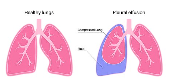
- The pleural space is a thin, fluid-filled cavity between the lungs and the chest wall.
- It prevents friction between the lungs and chest wall during breathing.
- Two membranes:
- Visceral pleura: Covers the lung surface.
- Parietal pleura: Lines the chest wall’s interior.
- Pleural fluid function:
- Acts as a lubricant.
- Reduces friction during inhalation and exhalation.
Functional Overview of the Lungs:
- Primary Function: Gas exchange.
- Oxygen (O₂) is absorbed from inhaled air.
- Carbon dioxide (CO₂) is released during exhalation.
- Gas exchange mainly occurs in the alveoli.
- Additional Functions:
- Metabolism of inhaled substances (e.g., volatile anesthetics, bronchodilators).
- Defense against airborne pathogens.
Lung Cell Types Mentioned:
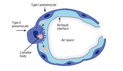
- Type I Pneumocytes:
- Thin, flat cells in the alveoli.
- Facilitate gas exchange (O₂ and CO₂ diffusion between air and blood).
- (Note: Other lung cell types like Type II pneumocytes and alveolar macrophages were not mentioned in this segment but are also key.)
Lung Cell Types and Their Functions (Expanded Explanation)
1 Type I Pneumocytes (Type I Alveolar Cells)
- Main Role: Facilitate gas exchange between alveoli and bloodstream.
- Structure: Thin, flat cells forming the alveolar wall.
- Function Details:
- Allow oxygen (O₂) to diffuse into capillaries.
- Allow carbon dioxide (CO₂) to diffuse out.
- Closely associated with capillaries surrounding alveoli.
2 Type II Pneumocytes (Type II Alveolar Cells)
- Main Role: Secrete pulmonary surfactant.
- Pulmonary Surfactant:
- A mixture of proteins and lipids.
- Reduces surface tension inside alveoli.
- Why it matters:
- Prevents alveolar collapse during exhalation.
- Helps maintain lung stability and ease of breathing.
- Additional Note:
- These cells can also regenerate Type I pneumocytes when needed.
Visual Aid Suggestion (From Transcript Context)
During the lecture, the speaker was asked to show a diagram of pneumocytes while explaining.
- A basic lung anatomy illustration showing:
- Alveolus structure.
- Type I pneumocytes lining the alveolar surface.
- Type II pneumocytes scattered among them, secreting surfactant.
- If you’d like, I can generate or sketch that diagram for you.
Type II (Expanded Clarification)
1 Core Function
- Secretion of Pulmonary Surfactant
- Composition: A mixture of proteins and lipids.
- Main Role:
- Reduces surface tension in the alveoli.
- Prevents alveolar collapse (especially during exhalation).
- Facilitates lung expansion during inhalation.
2 Why Surface Tension Matters
- Without surfactant:
- Alveoli would collapse due to the natural tension from the moist alveolar lining.
- Breathing would become extremely difficult or impossible.
3 Secondary Function
- Alveolar Repair and Regeneration
- Type II pneumocytes can transform into Type I pneumocytes.
- When Type I cells are damaged (due to infection, injury, etc.), Type II cells multiply and differentiate to restore the alveolar lining.
Final Segment Summary
1 Additional Lung Cell Types:
- Alveolar Macrophages
- Function:
- Engulf and remove foreign particles, allergens, pathogens, and dead cells via phagocytosis.
- Essential for lung defense and maintaining respiratory health.
- Function:
2 Fibrosis and Breathing Difficulties
- Pulmonary Fibrosis Mentioned:
- Defined as scarring or thickening of lung tissue, impairing gas exchange.
- Symptoms discussed:
- Dyspnea (difficulty breathing)
- Fatigue
- Weight loss
3 Naturopathy and Lung Health
- Naturopathic Approach Suggestions:
- Supportive role mentioned for chronic lung conditions such as fibrosis.
- Suggested methods:
- Diet modifications:
- Start with boiled foods for 3–4 days.
- Gradually introduce bland foods, then regular foods.
- Juices:
- Fresh vegetable and fruit juices suggested.
- Relaxation:
- Stress reduction and relaxation emphasized for patient well-being.
- Specific foods mentioned:
- Rice, carrots, papaya (suggested in soft and blended forms).
- Diet modifications:
Final Segment Summary: Patient Support & Naturopathic Management
1 Supporting Lung Function & Tissue Care
- Discussion Point:
- The speaker emphasizes tissue functioning and maintaining lung tissue health using naturopathic methods.
- References made to “ul neutral” (likely referring to pH balance or alkaline-neutral diets).
2 Patient Questions & Guidance
- Audience/Student Question:
- “Is it okay to give a patient with pressure…?” (Exact details unclear, possibly referring to blood pressure or pulmonary pressure management.)
- Speaker confirms: Yes, with appropriate naturopathic care.
3 Treatment Philosophy
- Key Message:
- The goal in naturopathy is to identify the root cause rather than only treating symptoms.
- Supportive care is emphasized:
- Help patients recognize triggering factors.
- Provide step-by-step dietary and lifestyle guidance.
4 Follow-Up and Ongoing Care
- Steps Mentioned:
- Ensure patients follow dietary recommendations strictly.
- Avoid sudden exposure to harmful factors (likely pollution, allergens, or cold air).
- Gradual process emphasized—no sudden shifts in care.
